Recommendations for proper handling of peripheral nerve biopsies (Lara & Kaspar Matiasek)
Peripheral nerve fibres are extremely prone to mechanical and chemical damage. Artefacts of excision and fixation may ruin the value of the biopsy. Therefore, a proper protocol has to be chosen with respect to several factors such as the time it takes until the probe is delivered to the histolab.
Key Points:
1. avoid stretching, crushing and drying of the nerve probe
2. if possible, immediate immersion in a fixative (mentioned below) is warranted
3. do not use formalin unless you are dealing with a peripheral nerve tumour
4. better no fixation than a wrong one
5. we can better handle autolysis than hyperfixation
6. envelop the nerve in wet gauze, don´t let it swim
...if in-house fixation is possible:
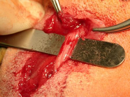
1. Remove abundant epineurial tissue softly.
2. Place single fascicles on a small sheet of paper in a longitudinal, gently stretched manner. This is to compensate for elastic properties of the nerve which, otherwise, cause a wavy fibre orientation that precludes proper logitudinal slices. It is a critical step, also, because contact to paper exsiccates the tissue quickly. To avoid a drying artefact, place the sheet immediately in the fixative, which is:
(stored) 2.5% glutaraldehyde
or
(freshly prepared) 4% paraformaldehyde in PBS.
Do not place the vessel in the refrigerator but let it go at room temperature. The time of immersion should not exeed 1 hour in aged animals, 2 hours in young animals. If you were able to habour very small fascicles 30 minutes fixation time is suitable, thereby, minimizing chemical artefacts. If separation of the fascicles was not possible prefixatively without application of strong forces, try it again after 15 minutes of incubation when the tissue is less susceptible to mechanical damage. Tear of the paper after several minutes so that the fixative can soake through the tissue from either side.
3. After fixation, the probe can be stored in Soerensen´s phosphate buffer or cacodylate buffer (0.2M; pH 7.4). Now (!), place the vessel in the fridge or send it to the lab.
...if in-house fixation is NOT possible:
1. Do not remove the epineurial tissue since it´s a evaporation barrier.
2. Wrap the nerve in a gauze moistured with isotonic HTK-, NaCl- or Ringer´s solution and place it in a plastic tube.
3. Best preservation is archieved when it´s sent together with cold-packs.
...if advanced diagnostic techniques have to be applied:
For most sophisticated methods, like in molecular pathological approaches, well defined protocols for tissue conservation might be indispensable. When genetic, biochemical, and molecular biological assays are necessary, please, contact us before taking a biopsy.